
What are dentists looking for in dental x-rays?
Dentists are primarily looking for cavities, as well as bone levels in your mouth to help diagnose if you need certain treatment planning. Periodontal disease can usually be diagnosed through x-rays as well.
Are dental x-rays safe?
Dental x-rays are very safe, contrary to popular belief. You can go through a full set of x-rays and still be well within the maximum radiation exposure per year. Due to modern technology, our x-ray heads are very localized in exposure, meaning all the x-rays we take are only exposing your oral cavity and nowhere else on your body.
Why are dental x-rays necessary?
Dentists consider x-rays one of their best diagnostic tools in helping patients maintain good oral health. X-rays help dentists diagnose relatively common disorders such as cavities, periodontal disease and infections. Without this ability to see inside a tooth and beneath the gums, more diseases would go unchecked, patients would experience more pain and discomfort and more teeth would be lost because proper treatment was not completed in time.
At Smiles by Design we have “new” digital radiography. The digital x-rays replace the ordinary dental film with a flat electronic pad or sensor. The x-rays hit the pad the same way they hit the film. But instead of developing the film in a dark room, the image is electronically sent directly to a computer where the image appears on the screen. Digital radiography reduces radiation by as much as 80 percent. Digital x-rays are also more detailed, meaning that even tiny changes that may not be noticeable with the naked eye or with the older style dental film can be caught earlier. The image is then automatically stored on the computer in your patient record.
Are there different types of dental x-rays?
There are a few different types of dental x-rays. When you come in for a new patient exam, we usually like to take a comprehensive set of x-rays where we get x-rays of every tooth in your mouth, as well as a panoramic x-ray that shows the entire mouth at once. After you've been established in a dental office and if you've had most of your treatment completed, routine x-rays typically consist of 4-6 bitewing x-rays taken once every a year.
The Major Dental Series of X-Rays
There are three major types of dental x-ray series: the initial full mouth series, the yearly bitewing series, and the Panoramic x-ray film.
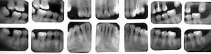
The Full Mouth Series (FMS)
This is an example of full mouth series we take in our office. It consists of 4 bitewing films, which are taken at an angle specifically to look for decay, and 14 periapical films, which are taken from other angles to show the tips of the roots and the supporting bone around the teeth. Not all full series look exactly like this one, but they all use some combination of bitewing and periapical x-rays to show a complete survey of the teeth and bones. We take a full mouth series on everyone over the age of 20 at the initial oral examination, and retake it again every 3 to 5 years.
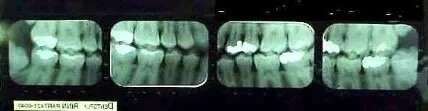
The Bite Wings
A bitewing series consists of either 2 or 4 films taken of the back teeth with the patient biting down so the films contain images of both the top and bottom teeth. A bitewing series is the minimum set of x-rays that most offices take to document the internal structure of the teeth and gums. In our office, we take 2 on children under the age of 12, and 4 on everyone older, supplemented by the other periapical films associated with a full series of X- rays if the patient is over the age of 20.
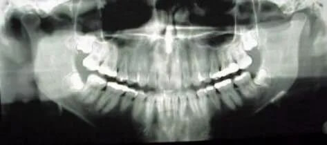
The Panorex or Panoramic
As you can see from the image above, the Panoramic is a large, single x-ray film that shows the entire bony structure of the teeth and face. It takes a much wider area than any intra oral film showing structures outside of their range including the sinuses, and the Temperomandibular Joints. It shows many pathological structures such as bony tumors and cysts, as well as the position of the wisdom teeth. They are quick and easy to take, and cost about the same as full series of intraoral films. In addition to medical and dental uses, panoramic films are especially good for forensic (legal) purposes.
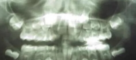
A Child’s Panoramic X-Ray
Above, you see a panoramic x-ray film of a child about 6 or 7 years old. You can see the adult teeth forming underneath the baby teeth. You see that the first molars (the 6-year molars) have erupted into place behind the last baby teeth in each arch, and so have the lower adult central incisors (you cannot see any lower baby centrals). But the upper adult central incisors have not yet erupted into position (you can see the upper baby centrals as little “stumps” sitting on top of the adult incisors). The adult second molars (the 12-year molars) are not expected to erupt until age 12. They are shown here only partly formed behind the fully erupted 6-year molars.